AdSense

Thursday, December 18, 2008

Blood Gas Analysis


Blood gas analysis
(BGA) is also known as arterial blood gas determination (ABG), and is considered a special test in the clinical laboratory. The commonly assayed parameters are partial pressure carbon dioxide (pCO2), hydrogen ion concentration (pH) and bicarbonate (HCO3). The determination of these substances helps in the evaluation of the acid-base status of a patient.

The following are certain precautions observed by medical technologists in the extraction of arterial blood for blood gas analysis.

1. The best specimen is arterial blood.

This is because arterial blood is more homogenous than venous blood. The blood could be collected in the following common arterial sites: the radial artery, the brachial artery, the femoral artery and the scalp artery.

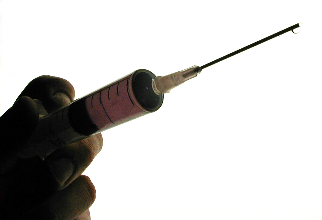
The most ideal anticoagulant is dry heparin, and the preferable syringe is a glass syringe. Recently, new receptacles are manufactured which are specifically for BGA.

2. Collect the specimen anaerobically (without air).

Your specimen should be covered at all times to prevent the escape of carbon dioxide to the air. It should be transported immediately to the testing laboratory. If it is left uncovered, unreliable results will be obtained which will lead to a wrong diagnosis by the Clinician/physician.

3. Preserve in crushed ice, if not tested immediately.

The low temperature has to be maintained. An increase in temperature would cause the gas to evaporate. It must also be preserved properly to obtain reliable results, aside from making sure that it is covered.


The body naturally maintains a state of balance called homeostasis. In the case of blood pH, this is done by the lungs and the kidneys which act as compensatory organs for one another. When the lungs are dysfunctional just like in respiratory diseases (emphysema, TB, chronic bronchitis, etc), the kidneys respond by excreting or retaining bicarbonate (HCO3).

On the other hand, when the kidneys are dysfunctional, the lung will respond by the increase retention or excretion of carbon dioxide (CO2). Through these physiologic processes, the critical pH (acidity and alkalinity) of blood is maintained at 7.35 to 7.45. Any slight variation from this pH, whether an increase or decrease, will lead to coma and eventually death. It is therefore imperative that the body maintains this slightly alkaline pH of blood for good health.

The following are acid-base conditions and the corresponding compensatory mechanisms :

Values : pH - decreased , PCO2 normal, HCO3 - decreased
Condition - metabolic acidosis
Compensatory mechanism - hyperventilation , increase excretion of CO2
decreased retention of CO2

Values: pH increased, HCO3 - increased , PCO2-normal
Condition - metabolic alkalosis
Compensatory mechanism - hypoventilation, decreased excretion of CO2
increased retention of CO2

Values : pH- increased , PCO2 - decreased , HCO3 -normal
Condition - respiratory alkalosis
Compensatory mechanism : increased retention of HCO3
decreased excretion of H+

Values: pH-decreased , PCO2 -increased, HCO3 -normal
Condition: respiratory acidosis
Compensatory mechanism : increased retention of HCO3,
increased excretion of H+

Clinical laboratory scientists or medical technologists solve for the pH of blood making use of the
Henderson-Hasselbalch Equation: (H & H equation). The formula for this is:

pH = 6.1 + log (HCO3)/DCO2

HCO = TCO2-DCO2

DCO2 = PCO2 X 0.031

Where: pH =indicates the acidity or alkalinity of a solution (hydrogen ion concentration.
HCO3 - bicarbonate
DCO2 - dissolved carbon dioxide
TCO2 - total carbon dioxide

Normal values are:

pH = 7.35-7.45

HCO3 = 22 - 26 mmol/L

PCO2 = 35 - 35 mmHg

TCO2 = 23-27 mmol/L

Arterial blood gas has very important clinical significances. It is crucial that the Medical Technologists know the precautions and perform the determination accurately. A well performed ABG signifies a patient well served.

Photo by NIOSH - Nat Inst for...


Reference:

Calbreath, Donald F. Fundamentals of Clinical Chemistry

Saturday, November 15, 2008

Review Questions at Clin Chem Reviewer

Click on the link to Clin Chem Reviewer and answer the review questions on BGA.

Photo by NIOSH - Nat Inst for...


Good luck.

Sunday, October 12, 2008

ANNOUNCEMENT

Photo by ccarlstead

THE FOLLOWING STUDENTS ARE REQUIRED TO REPORT ON TUESDAY;

OCTOBER 14, 2009, AT 10 AM.

VENUE: MT LAB

ID NOS.

1. 06-1718-758
2. 058205
3. 06-0330-934
4. 058501
5. 06-0132-304
6. 06-0322-615
7. 06-1861-244
8. 06-1028-725
9. 06-1062-179
10. 059289
11. 06-1720-892
12. 06-2060-419
13. 06-0323-469
14. 033776
15. 06-0466-976
16. 06-0142-221
17. 06-1801-109
18. 057131
19. 06-0935-794
20. 058866


COME IN COMPLETE UNIFORM AND BRING ALL NECESSARY PARAPHERNALIA.

DON'T FORGET YOUR CALCULATORS.

GOOD LUCK AND GOD BLESS.

VBG

Friday, September 26, 2008

How the body rids itself of fat

The body has a mechanism called homeostasis that keeps all the blood levels of substances, including fat, within normal levels in the bloodstream. This means that we need a certain amount of fat too.
This homeostatic function is done automatically if all of the organs in the body are functioning normally, and when the fat intake is not in excess.
Read more...

Understanding foodborne illness

Food borne illnesses go sometimes unnoticed because of the lack of understanding of its etiology or origin.
Understanding its root cause therefore is of utmost importance to people especially those in the food industry.
There are several sources of foodborne illnesses. Below are the most common among them:
Read more...


How to choose a multi-vitamin

Choosing the appropriate multi-vitamin for a person is important so that its maximum benefits could be obtained. TDM (Therapeutic Drug Monitoring) usually does this when time permits and the person is willing to undergo the procedure.
In therapeutic drug monitoring, drugs are administered to a certain person and observed for its therapeutic effect. The drug dosage is adjusted until such time that the dosage is therapeutic and neither sub-therapeutic nor toxic to the person.
Read more...


Healthy aspects of high fat food

Not all fatty foods are bad! This is because there are two types of fat: the High Density Lipoprotein (HDL) which is the good fat and the Low Density Lipoprotein, (LDL) which is the bad fat.
The good fats have several very important physiologic functions in the body: they act as constituents of bio-membranes (like phospholipids in the lungs, without it the lungs would not be able to contract and expand); they act as heat insulators and maintain warmth in the body; good cholesterol (a type of good fat) acts as the basic component of steroid hormones (progesterone, estrogen, testosterone). If this substance is absent what would happen to the full development of our secondary female and male sexual characteristics?
Read more...


Essential vitamins for pregnant women

The amount of essential vitamins needed by pregnant women is understandably increased than that of a woman who does not nurture a fetus in her womb.
Through the National Research Council of the FNBNAS (Food and Nutrition Board of the National Academy of Sciences), had issued an RDA (Recommended Daily Allowance) that could be used to determine the amount needed for proper nutrition.
Read more...

Monday, September 22, 2008

CHAMPIONS - PAMET NATIONAL QUIZ SHOW

We are the CHAMPIONS in the recently concluded
PAMET NATIONAL QUIZ SHOW.

This prestigious quiz show is being participated in by all Medical Technology Schools in the Philippines.

It feels great to be the CHAMPION.

Tuesday, September 9, 2008

Medical Technology Board Exam Results For September 2008


CONGRATULATIONS TO OUR NEW MEDICAL TECHOLOGY BOARD PASSERS RESULTS FOR SEPTEMBER 2008,
YOU MAY VISIT THE PRC SITE TO LEARN MORE.

Wednesday, September 3, 2008

FOR THE GROUP REPORTING QUESTIONS-PLS READ


FOR THE SUBMITTED QUESTIONS IN THE CLINICAL CHEMISTRY GROUP REPORTING, PLEASE CLICK ON THIS LINK TO VISIT THE SITE. THANKS.

Friday, July 18, 2008

COMMON LABORATORY APPARATUS


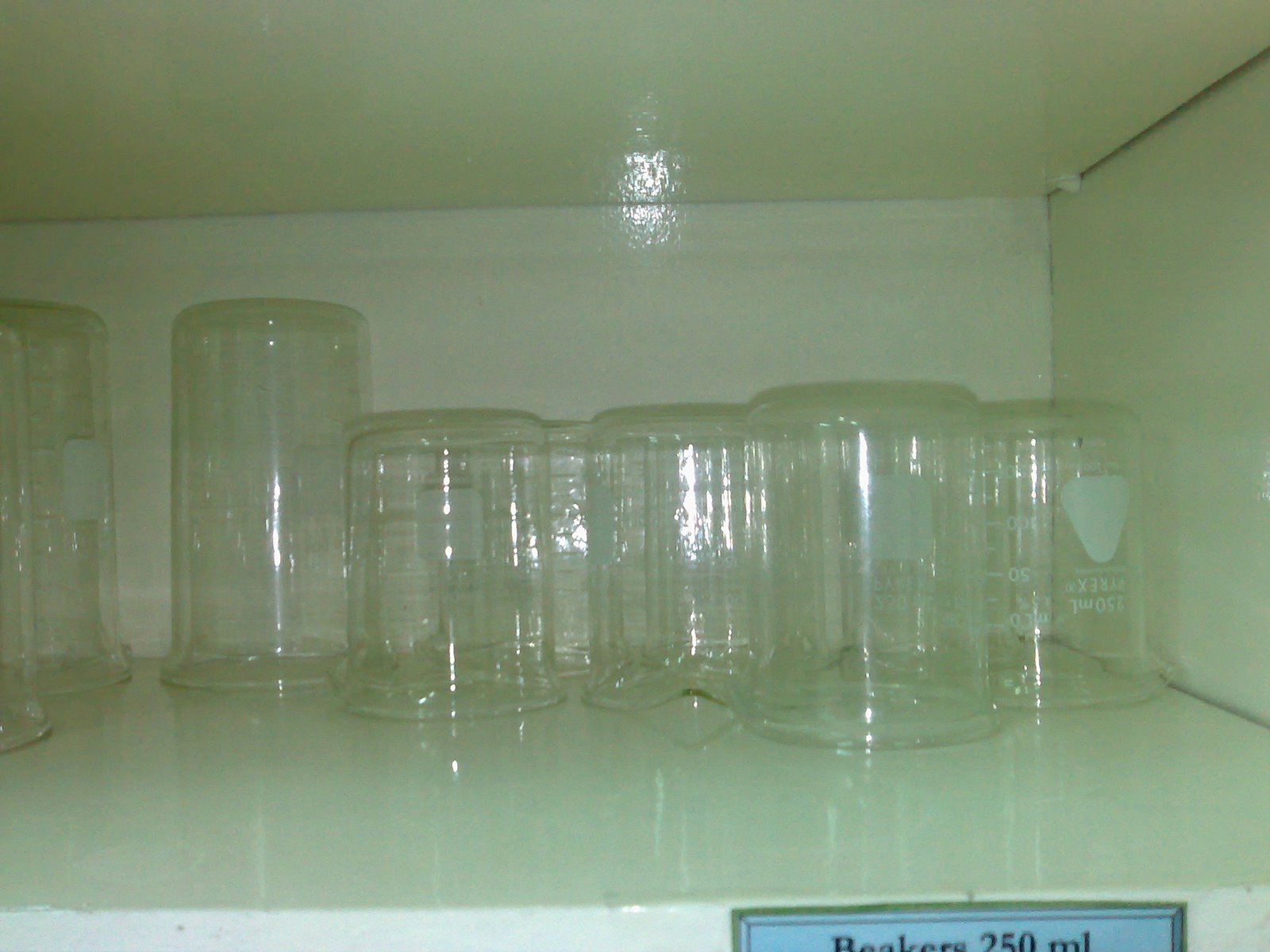
The beaker has many uses:

1. mixing solutions
2. measuring large amount of solutions
3. transferring solutions
4. reaction vessel

Friday, May 2, 2008

ORTHOTOLUIDINE METHOD FOR BLOOD GLUCOSE DETER MINATION


MANUAL PROCEDURE:

Precautions:

1. Patient should have fasted for 8-12 hours.
2. Unhemoylzed, non icteric and non turbid serum should be used to avoid interferences
with the procedure.
3. The reagent contains sodium azide, which is corrosive, so PPE should be worn.
4. The solution should be brought to a boil to ensure proper reaction.

PROCEDURE PROPER:

1. Prepare Three tubes labeled, SA (Sample)/ C (Control), ST (Standard) and RB (Reagent Blank).

2. To the SA/C tube add 0.1 ml of the test specimen/ Control specimen.

3. To the ST tube add 0.1 ml of the standard stock solution.

4. To the RB tube add 0.1 ml of distilled water.

5. Add 4 ml of glucose reagent to all tubes. Cap and mix by gentle inversion.

6. Boil all tubes at 100 degrees centigrade for 8 minutes.

7. Cool tubes in running tap water for about 2 minutes.

8. Read absorbance of solutions in the spectrophotometer with appropriate cuvets at 636 nanometers against the RB (Reagent Blank).

9. Compute for the Cu (Concentration of the Unknown making use of the formula:

Cu= Absorbance of Unknown(Au) multiplied by the Concentration of the
Standard(Cs) divided by the Absorbance of the Standard (As)

Adapt the unit of the Cs

Normal Values = 60 - 100 mg/dL
to convert values to mmol/L multiply values in mg/dL with 0.0555

Tuesday, April 29, 2008

FUNCTIONS OF LIPIDS

1. Heat insulator - retains heat in the body
2. Lung surfactant - phospholipid
3. Transport substance - lipoproteins
4. Basic nucleus for steroid hormones
5. Energy source - is a purer form that carbohydrate

For more information visit the link below:

associatedcontent.com

Monday, April 28, 2008

DIABETES MELLITUS

CLINICAL SIGNIFICANCE OF ELEVATED BLOOD GLUCOSE

Diabetes Mellitus

When the pancreas produces insufficient insulin or no insulin at all, Diabetes Mellitus results. This is because insulin is responsible in maintaining the normal blood glucose levels by several processes that allow the body’s tissues to absorb glucose and utilize it as energy or store it for future use.

Diabetes Mellitus is characterized by what is called the classical triad, or the three P's - polyuria, polydyipsia and polyphagia. Other symptoms also are hyperglycemia and glycosuria among others. This condition can affect several organs in the body.
For more information visit this site: MedicineNet.com


Image: FreeDigitalPhotos.net

Saturday, April 19, 2008

LABORATORY TESTS FOR GLUCOSE

Glucose is the most predominant carbohydrate and is the sugar tested in blood sugar
tests. There are certain preparations/precautions before an accurate laboratory test for glucose could be performed.

Whatever method is selected, the Medical Technologist must take the proper precautions in sample collection to prevent glucose utilization by leukocytes.

If the sample is left at room temperature, glucose in sample is reduced 10mg/dL/hour, therefore sample should be refrigerated within 30 minutes after extraction.

2 mg sodium fluoride per mL blood could be also added to prevent glycolysis for 24 hours.

Normal values:
Before meal: 90-130 mg/dL
After meal: < 180 mg/dL

There are Two General Methods for Glucose Detemination:

A. Chemical Methods - oxidation-reduction methods (ferric and cupric), condensation
methods
B. Enzymatic Methods - Hexokinase, Glucose Oxidase

Under the Chemical Methods: ( Alkalike copper reduction methods)

1. Folin-Wu - involves the conversion of phosphomolybdic acid to phosphomolybdenum blue

Reference value: 80-120 mg/dL

Precautions:
* Reagents should be freshly prepared
* Spectrophotometer: wavelength -520 nm
* Deprotienization should be done with the sample- removing of proteins from biologic fluids thru dilution

2 Nelson-Somogyi - involves the conversion of arsenomolybdic acid to arsenomolybdenum blue.
Spectrophotometer: 500-520 nm

3. Neocuproine - involves neocuproine reagent which produces a neocuproine complex.
• Spectrophotometer: 450 nm

4. Shaeffer-Hartmann-Somogyi - involves the use of iodine utilization as an indicator of the amount of glucose in the sample.
Spectrophotometer: 500-520 nm

5. Benedict’s

Reference value: 80-120 mg/dL
Precautions:
* Reagents should be freshly prepared

Chemical Methods (Alkaline Ferric Reduction Method)

1. Hagedorn-Jensen

Precautions:
* Traces of coagulum into the filtrate can cause increase in results.
* The stem of the funnel should have an ample amount of cotton.


Chemical Methods (Condensation Method)

1. Ortho-Toluidine
Gluconeer

Precautions:
* Non-icteric, non lipemic and unhemolyzed serum should be used.
* Patients should not be suffering from galactosemia
* Patients should not be under galactose load


www.freeimages.co.uk

B. Enzymatic Methods
1. Glucose Oxidase Method
2. Hexokinase Method

Reference Values:
* Fasting Serum Glucose: 70-110 mg/dL

* CSF:
Infant: 60-80 mg/dL
Adult: 40-70 mg/dL

* Urine (24 hours): 1-15 mg/dL

Precautions:
* Do not ingest the reagent, toxicity has not been established
* Spectrophotometer: 340 nm
* Use fresh, unhemolyzed serum. Plasma from tubes containing heparin, citrate,
oxalate or EDTA.
* Separate RBC from serum or plasma ASAP
* Glucose in whole blood is decreased 7% per hour.
* Turbid or icteric sera require sample blank

3. Glycosylated Hemoglobin
– HbA1c
Reference value: 4.0-6.0%
Precautions:
* Ample amount of blood- 5ml
* Apply pressure to the puncture site

FBS (Fasting Blood Sugar),the patient must have fasted for 8- 14 hours. This is because a non fasting patient will have an inaccurately increased value because of the effect of the ingested food.

The best anticoagulant in the specimen collection is sodium fluoride. This is because aside from acting as an anticoagulant, it also prevents glycolysis which can decrease inaccurately, the values obtained.

There are other tests like OGTT, (Oral Glucose Tolerance Tests), IGTT (Intravenous Glucose Tolerance Tests), RBS (Random Blood Sugar), 2HPPT ( 2- Hour Post Prandial Test)

ORAL GLUCOSE TOLERANCE TESTS

The oral glucose tolerance test (OGTT) measures the body's ability to metabolize glucose. This test is used to diagnose prediabetes and diabetes and to determine gestational diabetes in pregnant women.

Patient Preparation:

* Regular diet containing least 150 to 200 grams of carbohydrate per day is
observed for 3 days prior to the test.

* Medications should be referred to the doctor whether to stop or continue taking
them.

* Alcohol and cigarettes are not allowed at least 8 hours before blood collection.

* Strenuous exercise also is to be avoided at least 8 hours before blood collection.

* No unnecessary activities ( doing errands, manual labor) should be done during the
duration of the test.

* The patient is allowed water.

* If the fasting blood sugar is between 100-130 mg/dL, then OGTT is recommended.
Ranges higher than this, FBS is recommended.

* The patient should be ambulatory.

* The test should be performed in a hospital where a resident doctor can respond to
any emergencies.

* For polycystic ovary syndrome, a Glucose tolerance test screening is usually
performed at age 30 and above.

* Urine and fasting blood specimens should be tested first, before proceeding with
the test.

Friday, April 18, 2008

CARBOHYDRATES


Image from: Wikimedia.org
Carbohydrates are the main source of energy for the body. Sources of carbohydrates are rice, bread, pasta, corn, and many more. When ingested they are reduced to monosaccharide by the body's biochemical processes.

The most predominant monosaccharide is glucose. Glucose- also known as dextrose- is the sugar tested in the clinical laboratory when a person requests for a blood sugar test.

Blood glucose levels are maintained in the body at 60-100 mg/dL and this level is maintained through the body's homeostatic ability.

When the glucose levels increase the primary hormone that decreases this levels is insulin. On the other hand, the primary hormone that increases glucose levels when it is below normal, is glucagon. Both of these hormones are produced by the pancreas in the beta and alpha cells of the Islets of Langerhans, respectively.

Since blood glucose levels are affected by diet through the food that we eat, fasting is required before a test is performed( except in cases of RBS - Random Blood Sugar).

Fasting hours for 8-12 hours is done prior to the extraction of blood.

In the colorimetric method of glucose determination, visit the link below for full information and example:
Bioassaysys.com

Tuesday, April 15, 2008

LABORATORY MATHEMATICS

There are several methods of expressing concentrations:

1. Percent solutions - W/V (Weight over volume), W/W (weight over weight), V/V(volume over volume)

2. Normality - the gram equivalent weight of a solute per liter of solution.
3. Molarity - the gram molecular weight of a solute per lier of solution.
4. Molality - the gram molecular weight of solute per 1,000 gram of solvent.

Thursday, April 3, 2008

THYROID FUNCTION TESTS

THYROID FUNCTION TESTS

These are laboratory tests that are performed to determine thyroid function.

PRECAUTIONS IN SPECIMEN COLLECTION & HANDLING

•RED TOP OR YELLOW TOP CAN BE USED
•AVOID USE OF LIPEMIC & HEMOLYZED SERUM
•NEWBORNS- FILTER PAPER BLOT CAN BE USED
•FASTING- NOT NEEDED
•SAMPLES CAN BE REFRIGERATED-24 HRS.
•FROZEN FOR 30 DAYS (AVOID REPEATED THAWING)

BLOOD TESTS:

•Thyroid-Stimulating Hormone (TSH)
This is a significant marker for hypothyroidism. an increase in its concentration indicates hypothyroidism and a decrease indicates hyperthyroidism
Thyroxine (T4) and Triiodothyronine (T3)
increased concentrations indicate hyperthyroidism
decreased concentrations indicate hypothroidism
•Thyroxine-Binding Globulin. (TBG)
this is the major binding protein of thyroid hormones
Other tests:
rT3, T3U, FTI, FT3, FT4

Stimulation Tests

•-TRH STIMULATION TEST
TRH IS INJECTED INTO THE PATIENT AND THE RESPONSE OF THE PITUITARY GLAND IS OBSERVED.
•TSH STIMULATION TEST

TSH IS GIVEN TO THE PATIENT AND THEN THE REPONSE OF THE THYROID GLAND IS MEASURED.

THE CATECHOLAMINES

ADRENAL HORMONES
The CATECHOLAMINES are produced and metabolized in the ADRENAL MEDULLA.

The two most common catecholamines are:
1. EPINEPHRINE
2. NOREPINEPHRINE

These catecholamines are also called "EMERGENCY HORMONES" as they increase the ability of the body to respond to stressful situations.

They elevate blood pressure, increase glucose concentration in the blood stream and control a series of biochemical processes.

For more information about the topic visit the link below:

Become Healthy Now

Saturday, March 29, 2008

hCG -HUMAN CHORIONIC GONADOTROPIC HORMONE


hCG is the hormone being detected in most pregnancy tests. It is normally produced by the placenta during pregnancy and is also markedly elevated in pathologic conditions like seminoma, hydatidiform mole and teratoma, among others.

TOXICOLOGY


www.freeimages.co.uk

Divisions of Toxicology

1. Emergency - deals with overdosage of drugs
2. Drug Abuse Screening- deals with substances of abuse
3. Therapeutic Drug Monitoring - deals with drugs for therapeutic use
4. Forensic - deals with the medical -legal aspect
5. Industrial - deals with industrial products.

EMERGENCY TOXICOLOGY

SALICYLATE poisoning - is the most common cause of ET in children.
Salicylate/acetysalicylate - the active component of aspirin.

DRUG ABUSE SCREENING

Most common substances of abuse are cigarette and alcohol.

THERAPEUTIC DRUG MONITORING

The purpose of this is to establish the optimum concentration of the drug that could cure the patient.

FORENSIC

Prohibited drugs are usually involved in this.

INDUSTRIAL

pesticides and insecticides are the most common substances involved.

BLOOD GAS ANALYSIS OUTLINE

The determination of blood gas is considered a special test in the local setting.
pCO2, pH and HCO3 are usually assayed. The results help in the evaluation whether a patient has acidosis or alkalosis.

Conditions:

Values : pH - decreased , PCO2 normal, HCO3 - decreased
Condition - metabolic acidosis
Compensatory mechanism - hyperventilation , increase excretion of CO2
decreased retention of CO2

Values: pH increased, HCO3 - increased , PCO2-normal
Condition - metabolic alkalosis
Compensatory mechanism - hypoventilation, decreased excretion of CO2
increased retention of CO2

Values : pH- increased , PCO2 - decreased , HCO3 -normal
Condition - respiratory alkalosis
Compensatory mechanism : increased retention of HCO3
decreased excretion of H+

Values: pH-decreased , PCO2 -increased, HCO3 -normal
Condition: respiratory acidosis
Compensatory mechanism : increased retention of HCO3,
increased excretion of H+

Henderson=Hasselbalch Equation: (H & H)


pH = 6.1 + log (HCO3)/DCO2

HCO = TCO2-DCO2

DCO2 = PCO2 X 0.031


Where: pH =indicates the acidity or alkalinity of a solution (hydrogen ion concentration.
HCO3 - bicarbonate
DCO2 - dissolved carbon dioxide
TCO2 - total carbon dioxide

Sample Problems:

Based on the following lab results obtained,
1. What is the acid base status of the patient?
2. What further test would you recommend?
3. What may be the possible causes of the condition?
4. Show all computations.

laboratory data:

TCO2 = 20 mmol/L
PCO2 = 30 mmHg

For more on blood gas analysis, visit the site below:

labtests on line

Friday, March 28, 2008

TURBIDIMETRY

Turbidimetry measures the amount of light blocked by particles in a solution. The amount of light blocked is dependent upon its cross sectional area, its size and amount in the unknown solution. For more information click the link below:

Turbidimetry

ELECTROPHORESIS

Migration of charged particles or solutes in an electric field. The net charge of the molecule, their size and shape, the strength of the electric field and the support medium used, influence the rate and speed of distribution of the unknown particles.

'Electrophoresis' link';

Image from: http://en.wikipedia.org/?title=Gel_electrophoresis

EMISSION FLAME PHOTOMETRY (EFP)

• This method measures the concentration of an unknown solution by measuring the emitted flame from these solutions. Metal salts (Li,K,Na) are usually measured making use Of this method.


Image from this site, enter the LINK below for more information:

'Emission Flame Photometry! link';
Image from http://www.resonancepub.com/images/section701.gif

NEPHELOMETRY


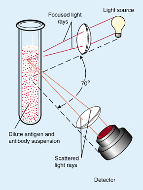
Image from: http://www.lib.mcg.edu/.../ch4/ch4img/nephelom.jpg
and http://www.gmi-inc.com/CliniLab/Beckman%20Immage.htm

Enter the LINK below to know more:

'Nephelometry' link';

NEPHELOMETRY

The light that is scattered by particles in the unknown solution is measured and made as a basis of determining its concentration.

Factors affecting the amount of light scattered are:

a.the wavelength used – dispersion of light is inversely proportional to the
wavelength.
b.the size of the particles
c.the amount of particles in solution
d.the cross sectional area of the particles
e.the source of light - monochromatic lights give more reliable readings because they
minimize sample heating and have more stable readings.

FLUOROMETRY

This method measures the fluorescence of substances in solution and make this as a basis of determining the unknown concentration. This is done by measuring the electromagnetic radiation absorption of excited atoms when they return to a higher energy level than their original levels. The wavelength used will be longer to the excitation wavelength.

Parts of a basic filter fluorometer:

1. Energy Source
2. Primary filter
3. cell or Sample holder
4. Attenuator
5. Secondary filter
6. Detector (Photomultiplier)
7. Readout

ENYZMOLOGY

The pancreatic enzymes:

1. lipase
2. amylase

The hepatic enzymes (TRANSCRIBE)

1. 5 nucleotidase
2. ALP
3. ALT
4. LAP
5. GGT
6. LDH
7. OCT
8. CHS - the only enzyme decreased in liver disease


Cardiac Enzymes (TRANSCRIBE)

1. CPK
2. AST
3. LDH

chi